Terminal-type sacral canal cysts often present as large leaks necessitating meticulous microsurgical suturing for resolution. Consequently, meticulous preoperative planning is paramount. Precise preoperative localization of the leak enables the surgeon to perform the closure procedure through a minimal bone window, facilitating a minimally invasive approach and preventing adhesion. Conversely, failure to identify the leak preoperatively necessitates a more extensive surgery involving the entire posterior wall of the sacral canal, resulting in significant trauma.
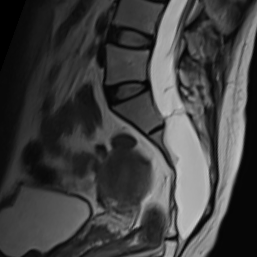
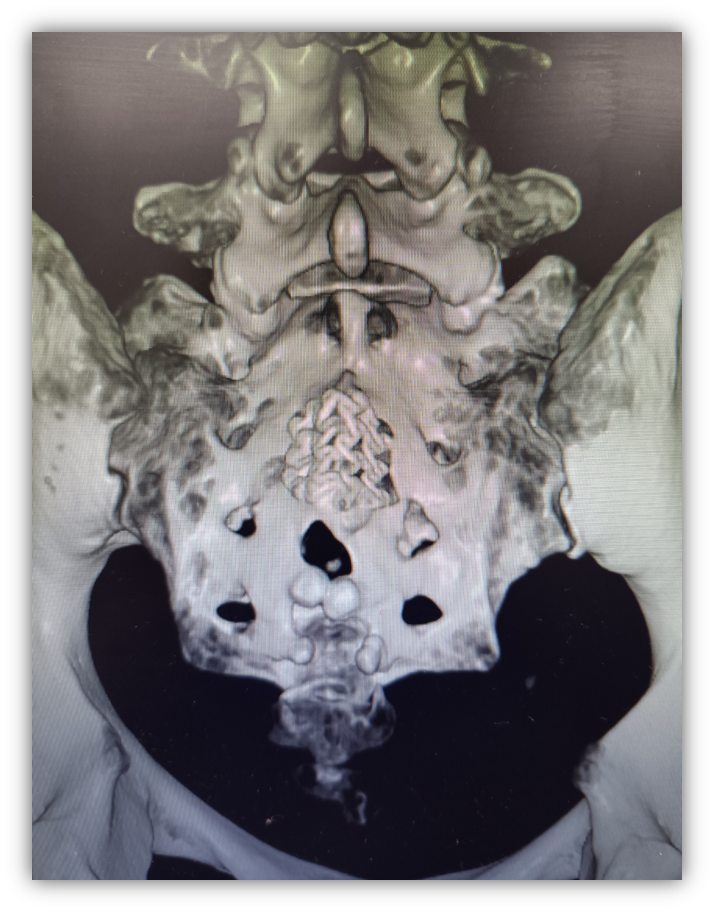
Thanks to preoperative imaging reconstruction and precise positioning, Dr. Xuesheng Zheng’s team pinpointed the leak in the right sacral plane and discovered that the bone in the left sacral posterior wall had been comprehensively eroded by a massive sacral cyst.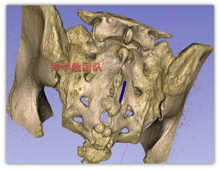
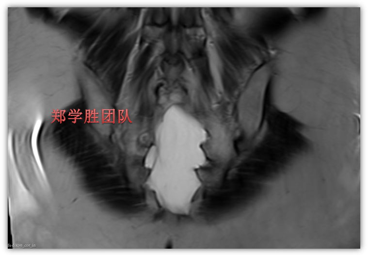
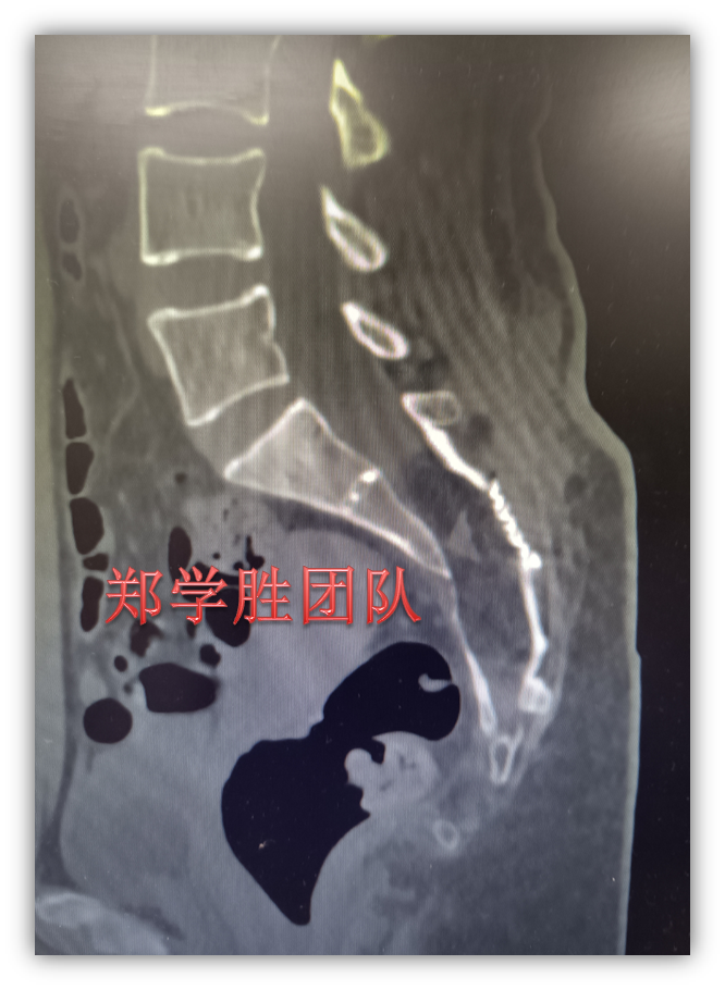
Guided by preoperative positioning, a small, approximately 1.2cm horizontal bone window was created on the right sacral canal during surgery to pinpoint and tightly seal the sacral cyst leakage, facilitating a smooth operation.
Postoperative MRI confirmed the successful sealing of the sacral cyst.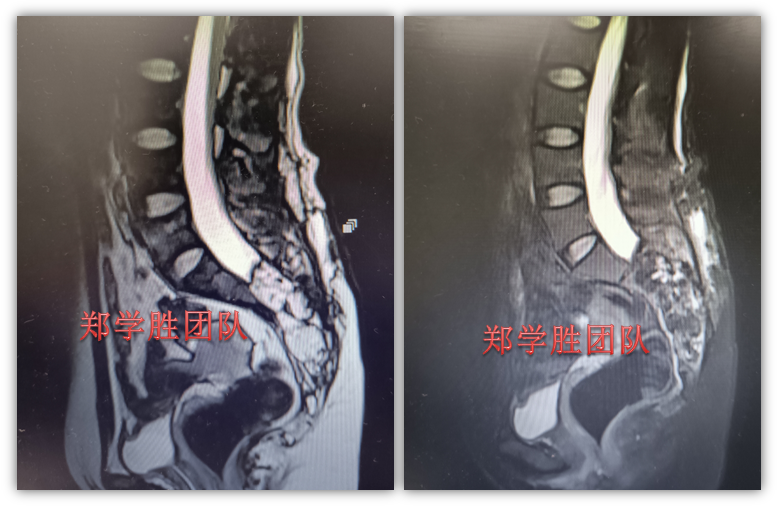
Chief Physician Xuesheng Zheng commented that giant sacral cysts severely compromise the sacrum bone, rendering it fragile and unable to support weight, thereby limiting patients’ ability to stand or walk. The patients are susceptible to sacral fractures, with bony fragments potentially damaging nerves. Therefore, surgeries for giant sacral cysts must prioritize bone protection. Accurate leak localization enables the use of a minimal laminectomy, thereby preserving the sacrum’s structural integrity. Some patients with giant sacral cysts experience severe bone erosion, seriously impacting their mobility, work capacity, and overall quality of life. Hence,the principles for giant sacral cyst surgical operation include minimal invasion, bone preservation, posterior wall repair, and adhesion prevention.
Please refer to: Repairing the posterior wall of sacral canal in Tarlov cyst operation procedure