A 25-year-old male patient was admitted to the hospital due to a growing mass in the lower right abdomen for two months, accompanied by stool weakness and constipation. Additionally, he experienced soreness and pain in the lumbosacral region, rendering him unable to stand or sit for prolonged periods and hindering his ability to maintain work performance.
Magnetic resonance imaging revealed a huge sacral cyst that had protruded through the sacral foramen into the pelvic cavity, forming a spherical cystic mass exceeding ten centimeters in size. Furthermore, the cyst had penetrated the posterior wall of the sacral canal, creating a conical cystic mass within the intermuscular space.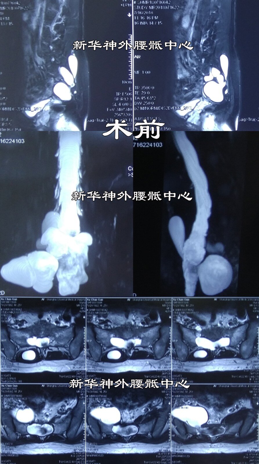
During surgery, it was discovered that the sacral cyst had a big leak, which was promptly sealed while ensuring the protection of transversing nerves.
Follow-up magnetic resonance imaging conducted within one year post-surgery demonstrated successful occlusion of the cyst with no signs of recurrence. To date, the patient has been under follow-up care for nearly two years.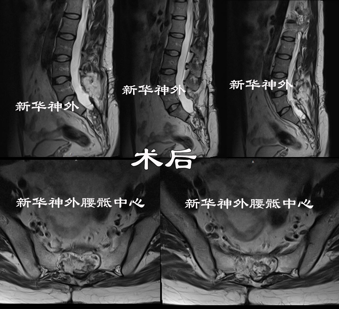
All symptoms subsided rapidly, and bowel movements recovered to normal. The patient resumed his work responsibilities three months after surgery.
Dr. Zheng Xuesheng emphasized that the prolonged exposure of sacral cysts to substantial static water pressure can lead to damage to the sacral vertebral body, which typically has a thickness of 3-4 centimeters. This damage allows the cyst to protrude into the pelvic cavity. Once in the pelvic cavity, the cyst loses the bone restriction, enabling rapid enlargement. In this particular case, the cyst affected bowel function within two months, causing stool weakness and constipation. If left untreated, it was anticipated that urinary and sexual function would suffer irreversible damage in the near future. Fortunately, the patient underwent timely surgery, resulting in swift postoperative recovery. Given his youthful and robust constitution, he was able to resume his duties within three months.